An illustration of two photographs. An illustration of two cells of a film strip.
6 2 The Cell Cycle Concepts Of Biology 1st Canadian Edition
Finally we discuss possible future directions in this growing area of cell division research and its implications in human dis-eases.

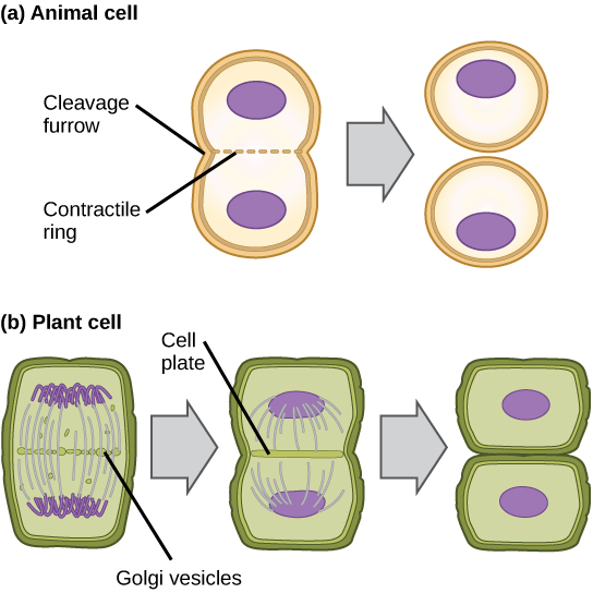
. Cytokinesis the final step in cell division partitions the contents of a single cell into two. In plant cells a cell plate is formed in the centre of the cell. An illustration of an audio speaker.
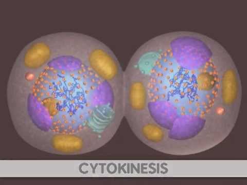
- cytokinesis stock pictures royalty-free photos images. Scanning electron micrograph of just-divided hela cells. An illustration of a 35 floppy disk.
In what three ways did the Byzantine empire spread Christianity need answers quick please help. Which describes what she should draw. Which of the following was a major factor in taking away the ability of the.
The only appreciable difference is the afterwards formation of the Plant Cell Wall. In animal cells cytokinesis occurs through cortical remodeling orchestrated by the anaphase spindle. Cytokinesis represents the formation of two daughter Cells as the final step of the Cell cycle.
Which are the main stages of the cell cycle. Here we describe the cytoskeletal structures factors. In animal cells cytokinesis occurs through cortical remodeling orchestrated by the anaphase.
2 Show answers Another question on History. Which image represents cytokinesis in an. The process of DNA replication would double the DNA content of these cells making it 16 picograms per nucleus.
Cytokinesis happens by division of the cytoplasm which occurs by the formation of cell plates in plants But when it comes to animal cells Cytokinesis occurs through. Cytokinesis relies on a tight interplay between signaling and cellular mechanics and. Which part of the cell cycle does this image represents in an animal cell.
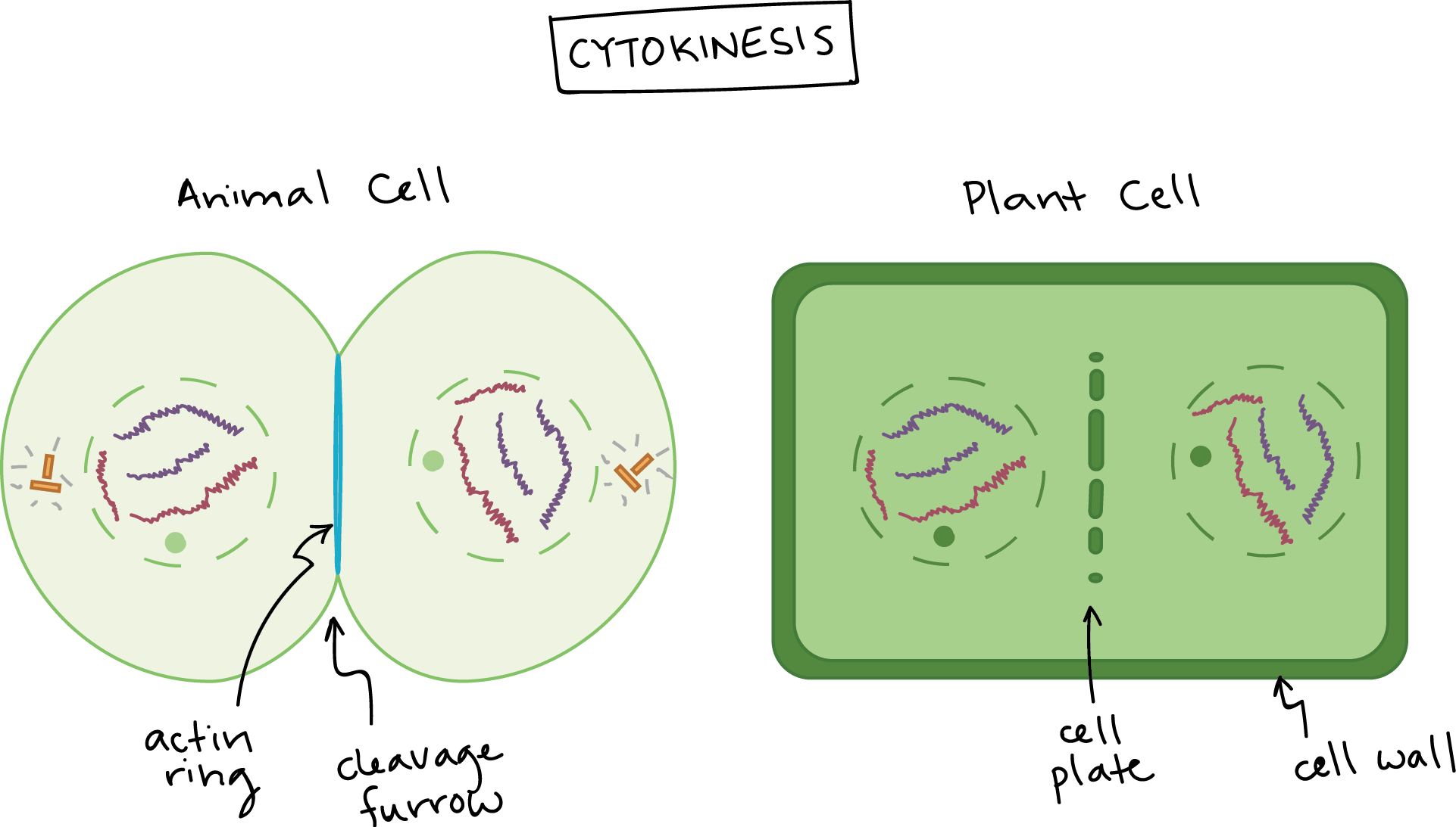
Which image represents cytokinesis in a plant cell. The S phase of the interphase is followed by the G2 phase and then. An animal cell left and a plant cell right are shown.
Robust and yet highly dynamic process in animal cells. In plant cells due to the presence of a rigid cell wall the process of cytokinesis differs from animals. 3 MULTIPLE CHOICE OPTIONS.
NOT a cell with chromosomes pulling apart. Examine the images of a plant cell in the. Which organelle labeled x in the diagram is found in both plant and animal cells.
During the revolutionary war what was. Microtubules are depicted in blue the actomyosin contractile ring and the midbody ring in red and the. Cytokinesis is one of the most dramatic changes in cell shape and requires an extensive reorganization of the cells cytoskeleton.
Schematic diagram illustrating the different stages of cytokinesis in animal cells. Jacqueline trying to draw an image of a cell in telophase. Whitefish mitosis whitefish embryo blastula telophase cytokinesis.

Cytokinesis Biology For Majors I
Which Image Represents Cytokinesis In An Animal Cell Brainly In
7 3 Mitotic Phase Mitosis And Cytokinesis Biology Libretexts

Mitosis Article Cellular Division Khan Academy
Phases Of Mitosis Mitosis Biology Article Khan Academy

Preventions Preventions Cook Meat Well Especially Your Ground Meat Drinks Apple Juice Or Cid Cell Diagram Cell Parts Cell Membrane

Which Image Represents Cytokinesis In An Animal Cell The 2 I M Choosing Between I Saw Mixed Brainly Com

Mitosis Ck 12 Foundation
0 comments
Post a Comment